More than 13,000 creatures scanned by scientists reveal stunning images of eggs inside a turtle, a bat’s ribcage and a rare snake eating a centipede
Scientists have scanned more than 13,000 creatures, finding eggs in a turtle, a bat’s ribcage and a rare rock snake that eats a centipede.
The project, called openVertebrate (oVert), was carried out by 18 institutions that spent six years scanning specimens from museums with the aim of providing researchers with useful data to make more discoveries about the animals.
The team used high-energy X-rays to see through the outside of each specimen, eliminating the need to dissect them, which could potentially destroy the creatures in the process.
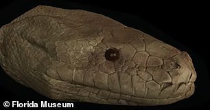
Scientists have already made discoveries using the scans, such as discovering that a rare rim rock crown snake was killed while trying to eat a centipede.
Meanwhile, it also revealed that a dinosaur called Spinosaurus, which was larger than Tyrannosaurus rex and long believed to have lived in water, was in fact a poor swimmer and would have stayed on land.
Scientists scanned 13,000 creatures, including amphibians, reptiles, fish and other mammals
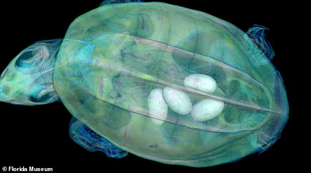
The 3D view of the turtle was difficult to obtain, so the scientists placed it on a raft to get a full scan
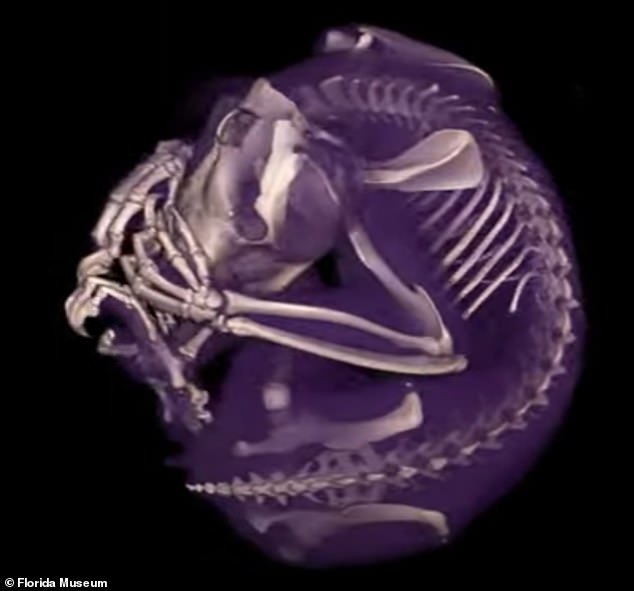
Scientists scanned the creatures for six years, from 2017 to 2023
Previously, this data could only be viewed by scientists who traveled to see the specimen or if it was sent to them on loan, but now researchers and the public can marvel at its inner workings anywhere in the world – and for free.
From 2017 through 2023, oVert project members captured CT scans representing more than half of all amphibians, reptiles, fish and mammals in the US
The research paper states that while they have been able to obtain the most specimens of amphibians and reptiles, they are unlikely to make more progress for birds and mammals due to the lack of available specimens in the US.
“Museums are constantly engaged in a balancing act,” says David Blackburn, principal investigator of the oVert project and curator of herpetology at the Florida Museum.
‘You want to protect specimens, but you also want people to use them.
“oVert is a way to reduce specimen wear and tear while increasing accessibility, and it is the next logical step in the mission of museum collections,” he said.
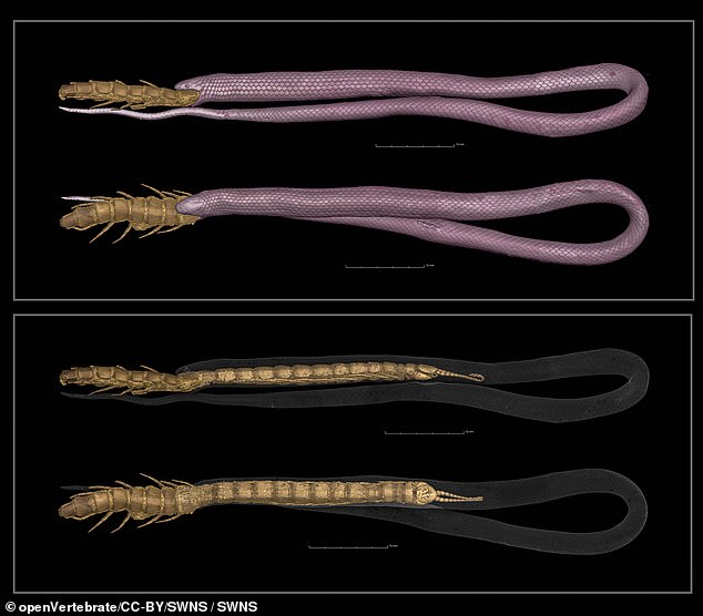
A rare rimstone snake died while trying to eat a centipede
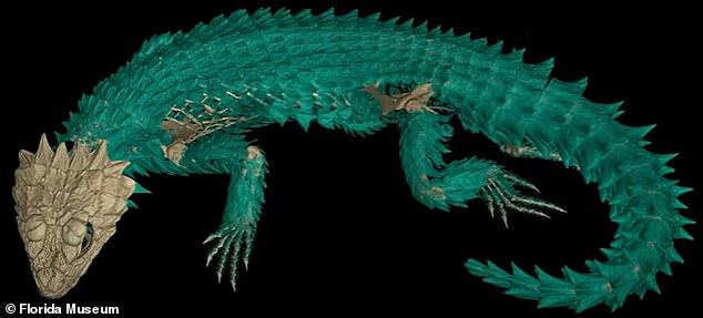
Scientists had to decide which specimens to use, and have still only scanned about half of the creatures still locked up in museums
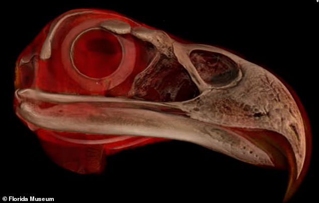
Scientists have managed to collect the most specimens of amphibians and reptiles, but don’t think they will make more progress for birds and mammals due to the lack of available specimens in the US.
In some cases, creating a 3D rendering of the creature was easier, by passing the specimen through the X-ray, but in other cases the project participants had to use their ingenuity to get a more complete picture.
The participants also had to decide which specimens to use, and although the original goal was to scan only samples preserved in ethyl alcohol, some specimens were too large for moisture preservation and the scientists did not want to leave them out.
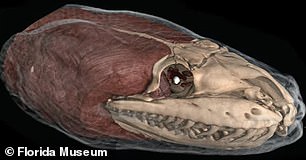
Most 3D models show before and after images of what the finished specimen looks like and the internal organs
For example, the Idaho Museum of Natural History wanted to make a digital model of a humpback whale, but it was too big to get a clear image with the scanner, so instead they took the skeleton apart and scanned and produced a 3D model. image of each bone.
When they were done, they reassembled the physical skeleton and the digital model.
These images are giving scientists a better understanding of how some of these creatures first functioned, including a model that showed that the fluid-filled canals in their pumpkin toad ears no longer functioned properly because they had become so small.
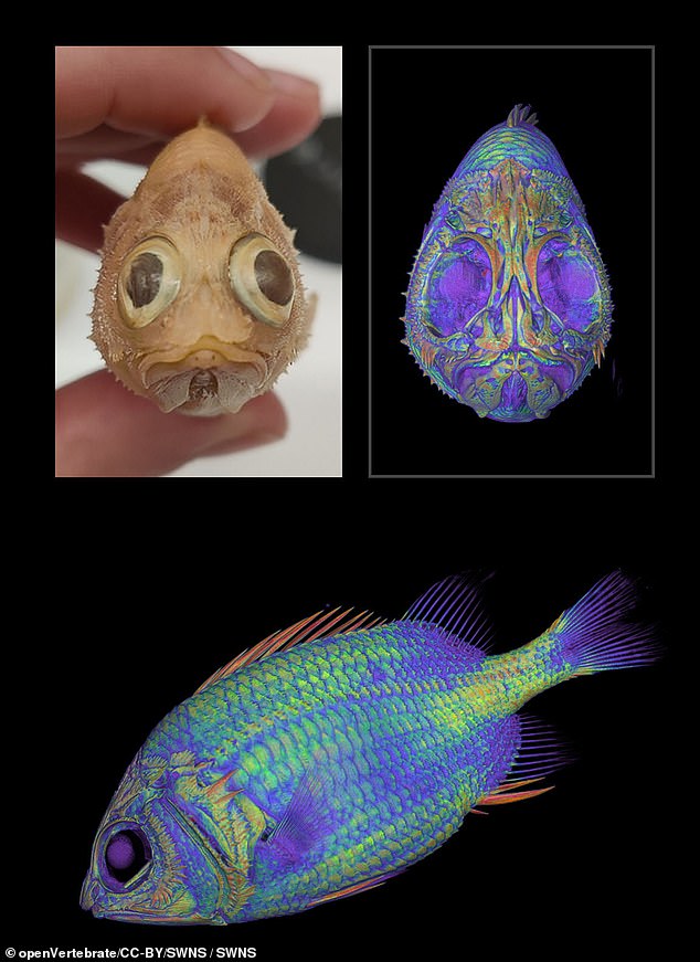
The oVert 3D data has been viewed and downloaded by scholars, undergraduate and graduate students, postdoctoral scholars, collection managers, K-12 teachers, and more
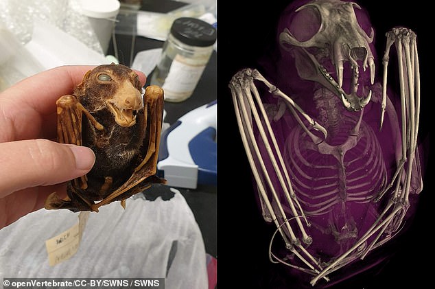
The oVert program is already used by over 3,700 people and the data has been viewed over a million times.
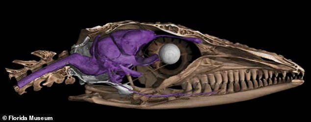
Scientists have already made countless discoveries because the 3D models show details they could not see before
The fluid channels produce electrical impulses in the brain that help the frog identify itself from top to bottom, but because they no longer work properly, the frogs crash land while jumping.
“You notice all kinds of things when you’re scanning,” says Edward Stanley, co-principal investigator of the oVert project and associate professor at the Florida Museum of Natural History.
Stanley was scanning spiny mice for the oVert project last year when he discovered that their tails were covered with internal bony plates called osteoderms, previously thought to be found only on armadillos.
“I study osteoderms, and through kismet or fate, I happened to be the one who scanned those particular specimens on that particular day and noticed something strange about their tails on the X-ray,” Stanley said.
‘That happens all the time. We found all kinds of strange, unexpected things.’
Blackburn said it’s important that people around the world can view these specimens because they don’t have to travel to collaborate, adding that in many ways it is unaffordable.”
He continued, “Now scientists, teachers, students and artists around the world have used this data remotely.”
As of March 2024, the program is already used by more than 3,700 people and the data generated by oVert has been viewed more than a million times.
The oVert 3D data was viewed and downloaded by scholars, undergraduate and graduate students, postdoctoral scholars, faculty, collections managers, K-12 teachers, exhibition staff and more, the oVert said research paper published in BioScience.
“It’s been a game-changer for my evolution unit,” Jennifer Broo, a high school teacher in Cincinnati, told the Florida Museum.
Broo told the outlet that her students can become less engaged when they know things are fake, but by using the oVert models, “you can teach concepts at an appropriate level while maintaining the authenticity of the science.”
