Inside the Human Heart: Shocking Video Reveals the Difference Between a Healthy Organ and One from a Cardiovascular Disease Patient
An incredible video takes you inside the human heart, showing the organ in more detail than ever before.
Experts in Britain and France used a new X-ray technique to capture anatomical structures down to 20 micrometres – half the width of a human hair.
In certain areas, imaging has been performed down to the cellular level, meaning that individual cells of the organ are visualized.
The clip compares two complete hearts from deceased adult donors: one healthy and one diseased.
While the healthy heart has a clearly defined shape, the diseased heart is rounder, with shriveled vessels and muscle fibers.
Healthy heart: Team’s technique captures the anatomical structure of the heart down to 20 micrometers – half the width of a human hair
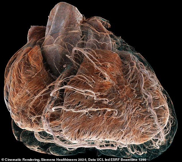
Diseased: This heart is from an 87-year-old white female donor with a history of ischemic heart disease
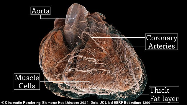
While the healthy heart has a clearly defined shape, the bad heart (pictured) is rounder, with shriveled vessels and muscle fibers
The images were taken by experts from University College London (UCL) and the European Synchrotron Radiation Facility (ESRF) in Grenoble, France.
“The atlas we have created in this study is like having Google Earth for the human heart,” said Professor Peter Lee from UCL’s Department of Mechanical Engineering.
‘This allows us to look at the entire organ on a global scale and then zoom in to street level to see cardiovascular features in unprecedented detail.’
The hearts come from two deceased patients. The healthy heart comes from a 63-year-old white male donor with no known heart problems.
The diseased heart came from an 87-year-old white female donor with a history of ischemic heart disease, a heart condition in which the heart is weakened by reduced blood flow.
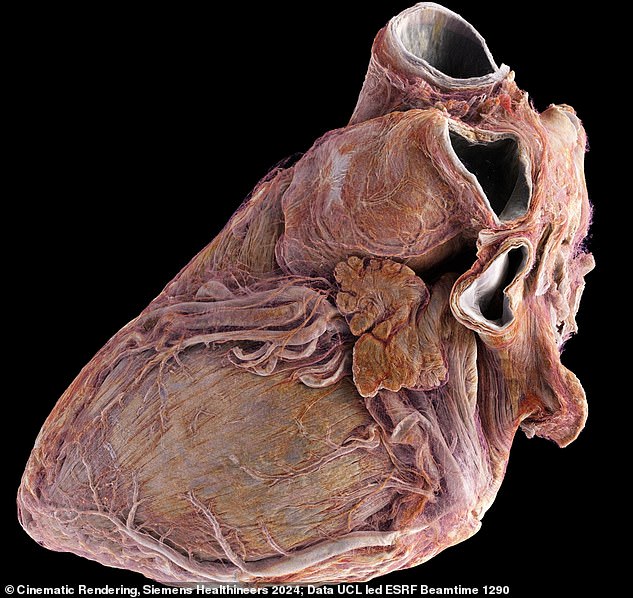
Healthy: Representation of a healthy male heart, showing external blood vessels and muscle fibers
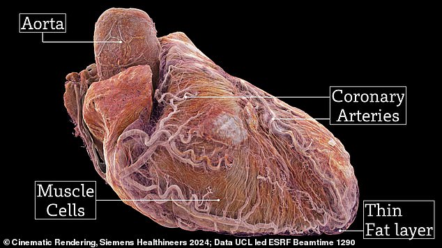
The healthy heart (pictured) came from a 63-year-old white male donor with no known heart problems
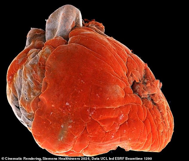
Sick: In addition to ischemic heart disease, the woman also had hypertension (high blood pressure) and atrial fibrillation (an irregular and often very fast heart rhythm)
Ischemic heart disease accounted for 8.9 million, or 16 percent, of deaths worldwide in 2019. This number has increased by more than two million since 2001.
The woman also suffered from hypertension (high blood pressure) and atrial fibrillation (an irregular and often very fast heart rhythm).
At the ESRF in France, the team used an X-ray technique called hierarchical phase-contrast tomography (HiP-CT) to image the hearts down to a scale of 20 micrometres.
“One of the biggest advantages of this technique is that it provides a full 3D image of the organ which is about 25 times better than that of a clinical CT scanner,” said Professor Lee.
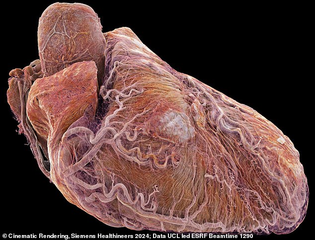
Healthy: The normal heart is a strong, hard-working pump, made of muscle tissue, about the size of a fist.
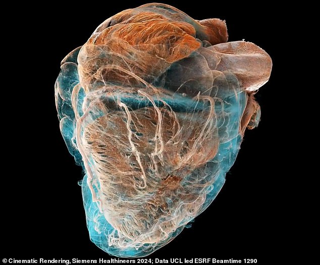
Sick: Ischemic heart disease was responsible for 8.9 million or 16 percent of deaths globally in 2019, a figure that had increased by more than two million since 2001
‘In addition, it can zoom in to the cellular level in selected areas, which is 250 times better. This allows us to achieve the same details as with a microscope, but without cutting the sample.
‘Because we can visualize entire organs in this way, details and connections come to light that we were previously unaware of.’
The researchers note that it is not without reason that the hearts involved come from deceased donors.
It is not possible to form an image of the heart of a living person in this way, because the radiation dose would be too high.
It may be some time before this technique is used routinely, as imaging each heart generates 10 terabytes of data – 1 million times more than a standard CT scan.
“The biggest limiting factor is processing the very large amount of data that HiP-CT produces,” said Paul Tafforeau of ESRF, the inventor of the technique.
Experts believe the images could be a tool to better understand cardiovascular diseases. Cardiovascular diseases are the collective name for various conditions that affect the heart or blood vessels and are the biggest cause of death worldwide.
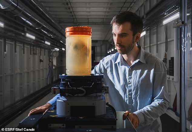
Joseph Brunet, UCL Senior Research Fellow and lead author, prepares to scan a human heart at the European Synchrotron Radiation Facility (ESRF)
‘We now have a way to determine the differences in the thickness of the tissue and fat layers between the outer surface of the heart and the protective sac around the heart, which could be relevant in the management of heart rhythm disorders,’ said Professor Andrew Cook, a cardiac anatomist from UCL’s Cardiovascular Sciences Institute.
The new study was published today in the journal Radiology.
A presentation of the research will take place at The Wonders, part of the UCL Festival of Engineering, on Friday 19 July at 7pm in the Bloomsbury Theatre.
