Breakthrough as US researchers ‘crack the autism code’
Researchers have developed a method for diagnosing autism that could save families years of uncertainty and allow treatment to begin sooner.
According to them, the new AI analysis can identify the genetic markers of autism through biological activity in the brain, with an accuracy of 89 to 95 percent.
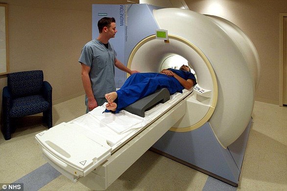
This new method starts with standard brain imaging using Magnetic Resonance Imaging (MRI). These scans are then reanalyzed using AI to detect the movement of proteins, nutrients and other processes in the brain that could indicate autism.
“Autism has traditionally been diagnosed behaviorally,” for example, through a person’s speech, as the medical team behind the trial noted. “But [it] has a strong genetic basis.’
A new method for diagnosing autism starts with standard brain mapping via magnetic resonance imaging, or MRI (pictured above), but reanalyzes those scans using AI to detect the movements of proteins, nutrients and other processes in the brain that indicate the condition.

Above, brain scan data comparing a control brain without autism (top row) to a brain containing deletions or duplications of genetic material associated with autism (bottom row). ‘Noise’ in this scan data was reduced on a 3D pixel-by-pixel basis using the ‘Z-score mapping’ method
According to the WHO, autism now affects one in 36 children. CDCwhich means that more than 90,000 children are born with this developmental disorder in the US each year.
But autism is notoriously difficult to recognize and the vast majority of children with the condition will not… diagnosed up to the age of five years and show clear behavioral characteristics.
Worse, the identification process usually involves years of uncertainty, dozens of hospital visits, and a battery of tests, including speech and language tests, observational interviews, and more. That can be stressful for children and families.
The researchers hope that the new diagnostic technique will soon allow doctors to pinpoint more specific genes responsible for autism by first uncovering the actual biological pathways through which autism changes the way a brain grows and functions.
As a spokesperson for a university behind the new method put it: the method ‘cracks the autism code’, although there is no information yet on when it will be widely used.
Dr. Shinjini Kunduassistant professor of radiology at Washington University in St. Louis, developed this new machine-learning AI, a mathematical brain modeling technique, when she was still a doctoral student researcher.
The method, called “transport-based morphometry” after the transport of biological material in the brain, focuses on identifying patterns associated with key bits of genetic code.
These sequences of genetic code, called copy number variations (CNVs), show that parts of the DNA have been deleted or duplicated. These changes have been linked to autism in previous research.
‘Some variations in the number of copies [CNVs] are known to be associated with autism,” said biomedical engineering professor Dr. Gustavo Rohde, who taught Dr. Kundu during her doctoral research.
“But their relationship to brain morphology — in other words, how different types of brain tissue, such as gray or white matter, are arranged in our brains — is not well understood,” said Dr. Rohde, who now teaches at the University of Virginia.
“Discovering the relationship between CNV and brain tissue morphology,” he explained, “is an important first step in understanding the biological basis of autism.”
Doctors Kundu and Rohde and their colleagues from the Department of Neurology at the University of California, San Francisco, published their results of developing this new method for autism identification in the journal Scientific progress.
Participants in the nonprofit Simons Variation in Individuals Project, a cohort of subjects with known autism-related genetic variations, provided key data used in the new study.
![Breakthrough as US researchers 'crack the autism code' 4 'Discover how CNV [deletion or duplication of genetic code]](https://nybreaking.com/wp-content/uploads/2024/08/1724955553_990_Breakthrough-as-US-researchers-crack-the-autism-code.jpg)
‘Discover how CNV [deletion or duplication of genetic code] “is related to the morphology of brain tissue,” said study co-author Dr. Gustavo Rohde, “an important first step in understanding the biological basis of autism.” The new method identifies these shifts in brain morphology
The researchers recruited their “control patients” from other medical or clinical settings based on their similarities to the Simons group (such as same age, gender, and nonverbal IQ), to limit variables that could confound their results.
According to Rohde, most machine learning methods that analyze medical data, such as MRI scans, do not include a mathematical model for the many biological processes hidden in that data.
Instead, previous AI models only looked for patterns to identify abnormalities or statistical anomalies in the health data of different patients.
However, Dr. Kundu’s “transport-based morphometry” could help researchers distinguish even more normal biological variations in brain structure — beyond the deletions or duplications associated with CNVs and autism.
Considering the reports that 90 percent of all medical data emerges from similar imaging, the team hopes this method can help extract new, useful information from old tools.
“Important discoveries can be made from such large amounts of data if we use more suitable mathematical models to extract such information,” said Dr. Rohde.
“We hope that the findings,” he added, “can point to brain regions and ultimately mechanisms that can be targeted for therapies.”

